
Why "Bone-on-Bone" Doesn't Tell the Whole Story
Sep 14, 2026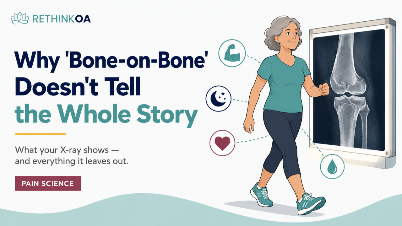
Few phrases in medicine land harder than this one: “You’re bone-on-bone.” People remember exactly where they were sitting when they heard it. For many, it instantly rewrites their future—no more hiking, no more tennis, surgery inevitable, downhill from here. The image it paints is visceral: two raw bones grinding against each other like unlubricated machine parts.
Here’s what I want you to know: “bone-on-bone” is a description of one finding on one image—not a verdict on your pain, your abilities, or your future. The phrase compresses an enormous amount of nuance into two scary words, and the science behind that nuance is genuinely good news. Let’s unpack what the phrase actually means, and everything it leaves out.
What “Bone-on-Bone” Actually Describes
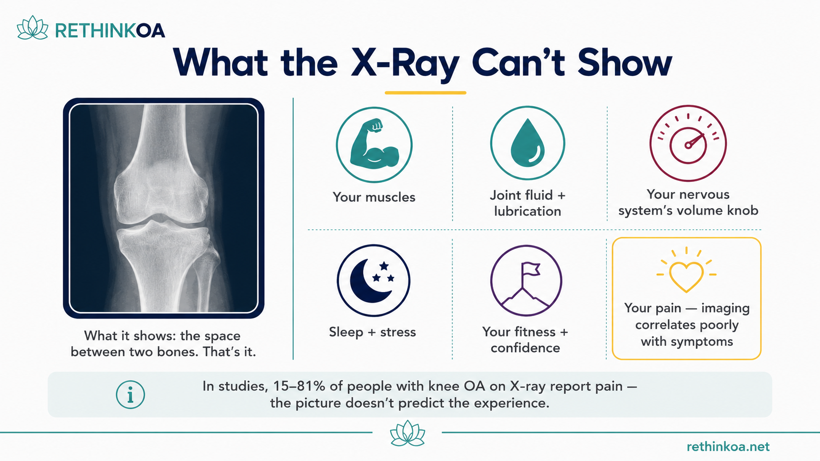
When a clinician says “bone-on-bone,” they typically mean that on your X-ray, the space between your femur and tibia looks narrowed—because the cartilage that occupies that space has thinned. A few important caveats hide inside that sentence. An X-ray is a two-dimensional shadow of a three-dimensional joint, taken in one position, on one day. It can’t show cartilage directly (cartilage is invisible on X-ray—its presence is inferred from the gap), it can’t show your muscles, and joint-space narrowing in one compartment says nothing about the cartilage that remains elsewhere in the joint.
Osteoarthritis is also a spectrum, not a switch. Having osteoarthritis—even advanced osteoarthritis on imaging—doesn’t mean the entire joint surface is in contact everywhere, all the time. And crucially, a joint is far more than the gap between two bones: it’s stabilized by muscle, bathed in lubricating synovial fluid, and continuously maintained by a body that adapts to how you use it.
The Picture and the Pain Are Two Different Things
If joint damage determined pain, people with the same X-rays would have the same symptoms. They don’t—not even close. A systematic review of the evidence found that among people whose X-rays show knee osteoarthritis, the proportion who actually have pain ranges from 15% to 81% across studies. Flip it around, and it’s equally striking: many people walking around pain-free today would show significant arthritis if you imaged them. A review of MRI studies found osteoarthritis-type changes—cartilage defects, bone spurs—are common in adults with no knee pain at all, and they become more common with age, like gray hair on the inside.
This is why pain is more than what shows up on an X-ray: pain is produced by a nervous system weighing many inputs—joint signals, yes, but also strength, sleep, stress, beliefs, and how threatening the situation seems. Two people can carry the same image in their medical charts and live in completely different bodies.
“Bone-on-Bone” Doesn’t Mean Improvement Is Off the Table
Perhaps the heaviest assumption baked into the phrase: if it’s bone-on-bone, nothing but surgery can help. The research says otherwise. In the landmark trial we’ve written about before, every participant had osteoarthritis severe enough to qualify for total knee replacement—and the group treated with exercise and education alone still improved meaningfully, with about two-thirds not proceeding to surgery within two years.
How can a knee improve if the cartilage doesn’t grow back? Because most of what makes a knee feel and function better was never about the cartilage gap:
- Stronger muscles absorb load and stabilize the joint—strength is highly trainable at any age and any X-ray grade
- A calmer nervous system turns down pain’s volume—through better sleep, less stress, understanding your pain, and graded activity
- Better fitness and weight support reduce the load carried across the joint with every step
- Restored confidence re-opens activities that fear had closed—often the biggest change in quality of life
None of those levers show up on an X-ray. All of them remain fully available to someone who’s been told they’re bone-on-bone.
Words Shape Outcomes—Choose the Accurate Ones
There’s a research-backed reason to care about the phrase itself. Threatening anatomical language—“bone-on-bone,” “wear and tear,” “crumbling”—can increase fear of movement, and fear predicts avoidance, deconditioning, and worse outcomes. In our clinical trial, simply teaching people more accurate ways to think about their osteoarthritis reduced fear of movement and self-imposed limitations, and increased physical activity. Words were the intervention.
So if “bone-on-bone” has been echoing in your head, try replacing it with something equally true and far more useful: “Part of my cartilage has thinned. The rest of my joint—muscle, fluid, bone, nervous system—is still working and still adaptable.” That’s not sugarcoating. That’s the fuller picture.
Final Thoughts
“Bone-on-bone” describes a picture. It doesn’t describe your pain—the correlation is famously loose. It doesn’t describe your trajectory—people with surgery-grade X-rays improve without surgery regularly. And it doesn’t describe your options—strength, fitness, sleep, understanding, and confidence remain open regardless of joint space. If those two words have been running your decisions, it may be time to hand the pen back to the rest of your body.
Rethinking what osteoarthritis means—accurately, based on evidence—is exactly what Rethink OA was built and clinically tested to do.
FAQ
What does “bone-on-bone” mean in knee osteoarthritis?
It’s informal shorthand for severe joint-space narrowing on an X-ray—meaning the cartilage between the femur and tibia has thinned enough that the bones appear close together in that view. It describes one imaging finding, not the state of the whole joint, and cartilage thinning is usually not uniform across the entire joint surface.
Does bone-on-bone always mean I need a knee replacement?
No. Surgery decisions are based on symptoms, function, and quality of life after a genuine trial of non-surgical care—not on the X-ray alone. In a randomized trial where everyone qualified for knee replacement, about two out of three people who received exercise and education had still not undergone surgery two years later. Many people with severe imaging findings manage well for years; some never need surgery.
Can a bone-on-bone knee get better without surgery?
Symptoms and function often can, even though cartilage doesn’t grow back. Research suggests improvement comes through trainable factors that don’t appear on X-rays: stronger supporting muscles, better fitness, weight management where relevant, improved sleep, reduced fear of movement, and a less sensitized nervous system. Exercise remains the first-line recommended treatment regardless of X-ray severity, appropriately dosed for you.
Why does my knee hurt so much if pain doesn’t match the X-ray?
Pain is real regardless of what imaging shows—it’s produced by a nervous system integrating joint signals with sleep, stress, strength, past experiences, and threat appraisal. Some people with mild imaging changes have severe pain, often involving a sensitized nervous system; others with severe changes have little pain. The mismatch doesn’t make your pain less real—it makes more of it potentially changeable.
Is it safe to exercise with a bone-on-bone knee?
For most people, yes—appropriately dosed exercise is recommended at every stage of knee osteoarthritis, and reviews of randomized trials show it doesn’t harm cartilage or increase adverse events. Start low, build gradually, and use symptom response as your guide. If you have significant health concerns or new severe symptoms, check with your clinician first—this article is general education, not individual medical advice.
About the Author
Melissa Boswell, PhD, is a bioengineer and digital health founder with nearly a decade of experience working in osteoarthritis, movement science, and human performance. She is the founder of Rethink OA, a clinically validated digital program developed from research conducted with collaborators at Stanford University and published in npj Digital Medicine. Her work focuses on helping people better understand pain, movement, and behavior change in osteoarthritis.

References
- Bedson J, Croft PR. The discordance between clinical and radiographic knee osteoarthritis: a systematic search and summary of the literature. BMC Musculoskelet Disord. 2008;9:116. https://bmcmusculoskeletdisord.biomedcentral.com/articles/10.1186/1471-2474-9-116
- Culvenor AG, et al. Prevalence of knee osteoarthritis features on magnetic resonance imaging in asymptomatic uninjured adults: a systematic review and meta-analysis. Br J Sports Med. 2019;53(20):1268-1278. https://bjsm.bmj.com/content/53/20/1268
- Skou ST, et al. A Randomized, Controlled Trial of Total Knee Replacement. N Engl J Med. 2015;373(17):1597-1606. https://www.nejm.org/doi/full/10.1056/NEJMoa1505467
- Skou ST, et al. Total knee replacement and non-surgical treatment of knee osteoarthritis: 2-year outcome from two parallel randomized controlled trials. Osteoarthritis Cartilage. 2018;26(9):1170-1180. https://pubmed.ncbi.nlm.nih.gov/29723634/
- Boswell MA, et al. A randomized clinical trial testing digital mindset intervention for knee osteoarthritis pain and activity improvement. npj Digital Medicine. 2024. https://www.nature.com/articles/s41746-024-01281-8
- National Institute of Arthritis and Musculoskeletal and Skin Diseases (NIAMS). Osteoarthritis. https://www.niams.nih.gov/health-topics/osteoarthritis
Move More. Fear Pain Less.
Learn how to reduce pain, stay active, and support your joints—starting today.